Dark Circles: Three Different Problems Wearing the Same Name
Concealer, then a second layer, then a third. Under-eye darkening is not one condition but three, and only one of them responds to sleep.
The routine is familiar enough that most women have stopped noticing it. Concealer, then a second layer at the inner corner, then a third when the light in the meeting room turns out to be unkind. By lunchtime the shadow has found its way back.
Dark circles are not one condition. They are at least three, and in a bathroom mirror they look almost identical.
One word covering three unrelated problems
The clinical term is periorbital hyperpigmentation, and the published literature is unusually frank about how loosely it is used. A comprehensive review of the condition calls it an ill-defined entity, and lists causes that have almost nothing in common with each other: inherited pigmentation, pigment left behind by eczema or an allergic reaction, swelling around the eye, unusually visible blood vessels, and shadowing thrown by the loss of support that comes with age.
A working classification did eventually emerge. The same review describes under-eye darkening sorted into four groups by what is actually producing the colour — pigmented, vascular, structural, and mixed. That last group matters more than it sounds, because most people arrive with a combination rather than a clean single type.
The pigmented type: brown, and genuinely pigment
This is excess melanin in the skin of the eyelid itself. It reads as a brown or brownish-black tone, and it holds its colour regardless of how rested you are or which way the light falls.
It has the strongest hereditary component of the three, and it is disproportionately common in deeper skin tones. It is also the type most often produced by something else — rubbing, an old bout of eczema, a reaction to a product used near the eye. That is pigment deposited after inflammation, and it behaves exactly like the marks left behind anywhere else on the face, which our guide to post-inflammatory hyperpigmentation sets out in detail.
The vascular type: not pigment at all
Here the colour is blue, purple or pink, and there is no excess melanin involved. The skin under the eye is the thinnest on the body, and the network of vessels sitting beneath it shows through — the same reason veins are visible at the wrist and not at the palm.
Anything that congests those vessels deepens the effect: poor sleep, crying, allergy, a long flight, a late night. This is the only type that genuinely responds to sleep, which is why the advice to sleep more works for some people and does nothing whatsoever for others.
The structural type: a shadow, not a stain
The third type has no colour of its own. As the face ages, fat pads under the eye lose volume and descend, the tear trough deepens, and the skin above it slackens. What appears is a shadow thrown by a contour — the eye socket’s own architecture becoming visible.
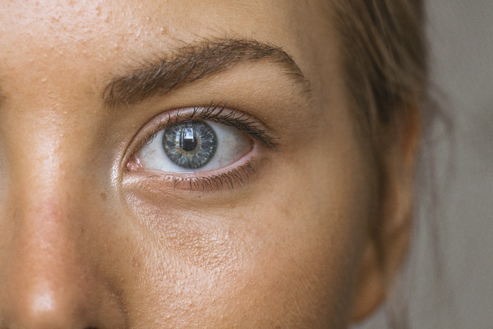
Two simple tests separate these in seconds, and the review names both. Gentle stretching of the lower eyelid skin is the first: true pigmentation keeps its colour when the skin is stretched, whereas a shadow improves or disappears entirely. And an increase in violet tone on stretching points to thin skin or vessels rather than pigment. You can perform the stretch test on yourself in front of a window, and it will tell you more than any product review.
| Pigmented | Vascular | Structural | |
|---|---|---|---|
| Colour | Brown to brownish-black | Blue, purple or pink | No colour — skin-toned shadow |
| What is producing it | Excess melanin in the eyelid skin | Vessels showing through thin skin | Contour: volume loss and laxity |
| Stretch test | Colour stays | Often deepens to violet | Improves or disappears |
| Changes with sleep | No | Yes, noticeably | Little |
| Worse in harsh overhead light | Unchanged | Unchanged | Markedly worse |
What the risk-factor research actually found
A cross-sectional study of 116 dermatology outpatients ran regression analysis across the usual suspects to see which ones held up. Family history, oral contraceptive use, the habit of rubbing the eyes, stress, diabetes and thyroid disorders all showed a significant association. Two stood out above the rest: rubbing the eyes, and prolonged screen exposure.
One finding cuts against expectation. In that model, prolonged sun exposure was not significantly associated with the condition. This does not make daily photoprotection optional — light is well established as an aggravator of pigment anywhere on the face, and our piece on why standard SPF leaves a gap for Indian skin explains why. It does suggest that for the under-eye area specifically, the mechanical habit of rubbing may be doing more damage than the sun is.
Which is a quietly uncomfortable finding, because rubbing is what people do when their eyes itch from allergy, from dry air-conditioned offices, and from screens held too close for too long. The habit both produces the pigment and is invisible to the person doing it.
Why most of the advice fails
Almost every remedy in circulation targets exactly one of the three types, and is then recommended to everybody.
Sleep and cold compresses address vascular congestion. Real effect, on the vascular type only. Caffeine serums constrict superficial vessels — again, vascular only, and temporarily. Brightening actives act on melanin, so they do nothing for a shadow or a vessel. Concealer covers colour but cannot fill a hollow; on the structural type it often deepens the shadow by settling into it.
Then there is the aggressive category. Strong acids and scrubs used near the eye on skin this thin do what over-treating does anywhere: they compromise the barrier and provoke inflammation, and inflammation in this area produces exactly the pigment being chased. Our article on barrier damage from over-exfoliation covers the mechanism, and the eyelid is the least forgiving place to test it.

What the evidence supports, by type
The review is clear that treatment has to follow the classification, because the types respond to different things.
For pigmented darkening, the evidence base sits with topical depigmenting agents — hydroquinone, kojic acid, azelaic acid and retinoic acid — with chemical peels and laser as procedural options. One reported protocol combined a bleaching preparation applied for six weeks beforehand with Q-switched ruby laser, and 15 of 18 patients showed excellent or good results after three to four sessions.
For vascular darkening, the target is the vessel rather than the pigment, and the review notes the 1064nm Nd:YAG laser as effective on both the vascular and pigmented components.
For structural darkening, no cream reaches the problem, because the problem is volume and contour. Options here are procedural and belong in a discussion about who is performing the injection rather than what is being injected — the tear trough is among the least forgiving areas on the face.
Applying the right treatment to the wrong type is the single most common reason people conclude that nothing works on dark circles.
How a clinical assessment approaches under-eye darkening
An expert assessment starts by establishing which type is present, and in what proportion — because mixed presentations are the norm and each component needs a different answer. The stretch test and examination under magnification separate pigment from shadow from vessel; a Wood’s lamp examination can further distinguish pigment sitting in the upper layers from pigment sitting deeper, which changes what is realistic.
It then looks for the drivers the research identifies rather than treating the surface: allergic or atopic irritation causing the rubbing, thyroid or metabolic factors, contraceptive use, sleep and screen patterns. It considers phototype, since deeper skin both pigments more readily and reacts more readily to procedures — a point our overview of where facial pigmentation comes from returns to repeatedly.
Only then does a personalised, clinically supervised plan follow: the aggravating habit addressed first, evidence-based topicals matched to the actual type, and any procedure introduced with precision and conservative settings on skin this delicate. The realistic goal is a meaningful reduction in how much the area draws the eye — and, for most people, the confidence of not reaching for a third layer of concealer by lunchtime.
Frequently asked questions
Will sleeping more get rid of my dark circles?
Only if yours are the vascular type, where congested vessels beneath thin skin are producing a blue or purple tone. Sleep has little effect on pigmented darkening and none at all on a shadow cast by contour, which is why the advice works dramatically for some people and not at all for others.
How can I tell which type I have at home?
Gently stretch the skin of the lower eyelid sideways and watch what happens to the colour. Pigment holds its colour, a structural shadow improves or vanishes, and a vascular component often deepens to violet. It is not a substitute for examination under magnification, but it is a genuinely useful first sort.
Are under-eye creams worth using?
A brightening formulation can help genuine pigmentation with consistent use over months. It cannot fill a hollow or hide a vessel. Before buying anything, work out which type you are treating — otherwise you are testing a pigment product against a problem that is not made of pigment.
Why do my dark circles look worse on video calls?
Overhead and front-on lighting exaggerates contour, so a structural shadow deepens sharply under it. Long screen sessions also feature in the risk-factor research alongside eye rubbing, and the two tend to travel together — dry, tired eyes get rubbed, and rubbing deposits pigment.
This article is for general education and is not a substitute for an in-person consultation. Treatment suitability depends on your medical history, skin type and current medication. Always speak with a qualified practitioner before starting or stopping any treatment.